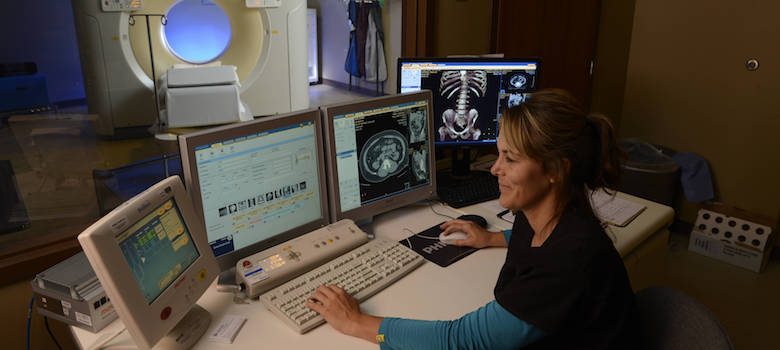
Caldwell Medical Center is proud to offer high-tech medical imaging and advanced diagnostic equipment. We strive to provide our patients with state-of-the-art technology right here at home, allowing our team of experienced medical personnel to deliver better radiology services for you and your family.
Our Canon Vantage Fortian MRI machine is the latest in advanced medical imaging technology! The Vantage Fortian offers higher patient comfort with a design created specifically for faster exam times, more comfortable positioning, an MR theater feature to help relax patients with a virtual, immersive experience, and Pianissimo Zen technology that offers whisper quiet scanning for a more calming feel.
This top-of-the-line equipment also provides your medical team with advanced imagery, offering clear, sharp and distinct images using 2D, 3D, Fast 3D and parallel imaging technology for reduced distortion, motion correction and advanced diagnosis capabilities.
Comprehensive diagnostic services provide improved imagery, faster exam times and less discomfort for our patients. From mammography, MRI and ultrasounds to bone density screenings and much more, Caldwell Medical Center’s imaging Department has the expertise and technology to meet all your diagnostic needs – right here, close to home.
Our Diagnostic Services Include:
Mammography:
The most effective method of screening for breast cancer, this type of imaging uses a low dose of x-rays to examine the breast for abnormalities.
Caldwell Medical now offers Genius™ 3D Mammography™! Click here to learn more >>
CT Scan:
Computed tomography (CT of CAT) scan allows doctors to see insider your body. It uses a combination of X-rays and a computer to create pictures of your organs, bones, and other issues. It shows more detail than regular X-ray services. You can get a CT scan on any part of your body.
MRI:
An MRI scan uses a large magnet, radio waves, and a computer to create a detailed, cross-sectional image of internal organs and structures. An MRI scan differs from CT scans and X-rays, as it does not use potentially harmful ionizing radiation. MRI scans are some of the most common forms of radiology services.
Ultrasound:
Diagnostic ultrasound, also called sonography or diagnostic medical sonography, is an imaging method that uses high-frequency sound waves to produce images of structures within your body. The images can provide valuable information for diagnosing and treating a variety of diseases and conditions.
Diagnostic X-ray:
X-ray services are used to diagnose bone abnormalities. Some internal organs are also imaged by using barium or an IV contrast.
Nuclear Medicine:
Nuclear medicine imaging uses small amounts of radioactive materials called radiotracers that are typically injected into the bloodstream, inhaled or swallowed. Nuclear medicine imaging provides unique information that often cannot be obtained using other imaging procedures and offers the potential to identify the disease in its earliest stages.
Bone Densitometry:
Also called dual-energy x-ray absorptiometry, DEXA or DXA, uses a very small dose of ionizing radiation to produce pictures of the inside of the body (usually the lower spine and hips) to measure bone loss. It is commonly used to diagnose osteoporosis and to assess an individual’s risk for developing osteoporotic fractures. DXA is simple, quick, and noninvasive. It’s also the most commonly used and the most standard method for diagnosing osteoporosis.
Looking for Diagnostic Services? Reach Out
You can find a provider with Caldwell Medical Center today. We look forward to hearing from you.


